Ultrasonic Diagnostic Equipment: Practical Applications and Techniques 2026
Ultrasonic diagnostic equipment plays a pivotal role in modern medical and veterinary practices. This guide aims to provide laboratory professionals with a practical understanding of how to use these devices effectively in 2026. This includes detailed applications, techniques, and best practices to optimize workflows.
What is it used for in 2026
In 2026, ultrasonic diagnostic equipment is primarily used for various imaging purposes across different fields, particularly in medicine and veterinary sciences. Its applications include:
- Medical Imaging: Used for diagnosing conditions in obstetrics, cardiology, and internal medicine.
- Veterinary Applications: Essential for prenatal examinations and other diagnostic procedures in animals.
- Research: Useful in laboratory settings for studies involving soft tissue analysis and vascular assessments.
History and evolution of the technology
The evolution of ultrasonic diagnostic technology dates back to the early 20th century when ultrasound waves were first explored for industrial applications. Over the decades, advancements in technology led to the development of portable and more sophisticated ultrasonic diagnostic devices. In recent years, the integration of digital imaging and software improvements has significantly enhanced the resolution and usability of these tools, making them indispensable in both medical and veterinary fields.
How to use it step by step
Using ultrasonic diagnostic equipment effectively involves several key steps:
- Preparation: Ensure that the equipment is charged and calibrated. Select the appropriate probe for the specific examination.
- Patient Preparation: For human patients, ensure they are in a suitable position. For veterinary patients, ensure calmness and stability.
- Conduct the Examination: Apply gel to the probe to facilitate sound wave transmission. Move the probe across the area of interest while monitoring the display for clear images.
- Image Capture: Use the device's software features to save images and generate reports as necessary.
- Post-Examination: Clean the equipment and document findings accurately.
Best techniques and protocols
To ensure optimal results when using ultrasonic diagnostic devices, consider the following techniques and protocols:
- Proper Gel Application: Use an adequate amount of gel to ensure clear imaging.
- Adjust Settings: Customize settings such as gain, depth, and frequency based on the specific examination type.
- Regular Training: Regular training sessions for staff to keep up-to-date with the latest protocols and features.
Practical applications by laboratory type
Different laboratories utilize ultrasonic diagnostic equipment in various ways:
- Medical Laboratories: Commonly used for obstetric imaging, cardiac assessments, and abdominal studies.
- Veterinary Clinics: Essential for evaluating pregnancy in animals and diagnosing health issues.
- Research Laboratories: Employed in studies related to vascular imaging and soft tissue analysis.
Regulations, standards and certifications
In 2026, compliance with regulations and standards is crucial for the operation of ultrasonic diagnostic equipment. Laboratories must adhere to:
- FDA Regulations: Ensure devices are approved for medical use.
- ISO Standards: Follow international standards for quality and safety.
- Equipment Certifications: Regular checks and certifications to guarantee performance and safety.
Comparison with alternative technologies
When comparing ultrasonic diagnostic equipment with alternative imaging technologies like MRI or CT scans, several factors come into play:
- Cost: Ultrasonic equipment is generally more cost-effective with lower operational costs.
- Safety: Non-invasive and does not involve radiation, making it safer for patients.
- Portability: Many ultrasonic devices are portable, allowing for use in various settings.
Comparison of available models
| Model | Best for | Key specs | Recommended use case |
|---|---|---|---|
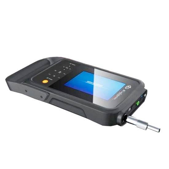
| YR05146 | Veterinary imaging | Lightweight, high-resolution display, 1024 image storage | Portable veterinary examinations |
| YR05147 | Obstetric and gynecological imaging | 15-inch LED monitor, 2D/3D imaging | Comprehensive women's health assessments |
| YR05154 | General diagnostic purposes | Single convex probe, versatile software | Routine examinations and diagnostics |
| YR05154-1 | Mobile diagnostics | Tablet integration, compact design | On-the-go examinations in various settings |
| YR05155 | Soft tissue imaging | Single linear probe, user-friendly software | Focused imaging of soft tissues |
| YR05155-1 | Advanced imaging solutions | Tablet integration, multiple functionalities | Complex examinations and diagnostic needs |
Common mistakes and how to avoid them
Here are some common mistakes professionals make when using ultrasonic diagnostic equipment and tips to avoid them:
- Inadequate Patient Preparation: Always ensure the patient is properly positioned and informed about the procedure.
- Insufficient Gel Application: Apply enough gel for optimal imaging quality.
- Ignoring Device Calibration: Regularly calibrate the device to maintain accuracy.
Maintenance, calibration and good practices 2026
Proper maintenance and calibration are essential for the longevity and accuracy of ultrasonic diagnostic equipment:
- Regular Cleaning: Clean the probes and equipment after each use to prevent contamination.
- Scheduled Calibration: Adhere to a strict calibration schedule as per manufacturer recommendations.
- Training Staff: Ensure all users are trained in good practices and maintenance routines.
Cost-benefit analysis 2026
When considering the investment in ultrasonic diagnostic equipment, evaluate the following cost-benefit aspects:
- Initial Costs: Compare the purchase price with budget constraints.
- Operational Costs: Consider ongoing costs such as maintenance, consumables, and staff training.
- Return on Investment: Assess how the enhanced diagnostic capabilities can lead to improved patient outcomes and workflow efficiency.
Frequently asked questions
What should I consider when choosing ultrasonic diagnostic equipment?
Consider your specific needs, such as the types of examinations you will perform, portability, and budget constraints. Evaluate the features and specifications that match those needs.
How often should ultrasonic equipment be calibrated?
Ultrasonic equipment should be calibrated regularly, typically every 6 to 12 months, or as recommended by the manufacturer to ensure accurate results.
Can ultrasonic diagnostic equipment be used in emergency situations?
Yes, many ultrasonic devices are portable and can be used in emergency situations for rapid assessments, making them valuable in urgent care settings.
What is the importance of using gel during examinations?
Gel is crucial as it eliminates air pockets between the probe and the skin, allowing for better transmission of sound waves and clearer images.
How can I ensure the best image quality with ultrasonic equipment?
To ensure the best image quality, use appropriate gel, adjust settings based on the examination type, and maintain the equipment properly.
What types of software are available with ultrasonic diagnostic devices?
Many devices come with integrated software for various applications, including obstetric, cardiac, and general veterinary assessments, as well as image management features.
How can I request a quote for ultrasonic diagnostic equipment?
You can easily request a quote by reaching out through our contact channels for detailed product information and pricing.
If you are looking for a fusion of innovation and quality, you have come to the right place. At Kalstein, we offer you the luxury of exploring our exclusive catalog of laboratory equipment. We manufacture every device to the highest standards of excellence. Our intuitive and seamless online purchasing channels are designed for your convenience, securing the most competitive prices. Hesitate no longer — we bring science to life, it is time to become part of our community.